Ear correction (otoplasty – correction of protruding ears)

In most cultures, the human ear holds no particular aesthetic significance. People don’t typically speak of beautiful ears, or of youthful or old ears. In contrast, we consider such attributes completely normal when describing the face or eyelids.
Before and After Photos

What are protruding ears? – Why can ears appear to stick out?

Most people cannot describe what makes an ear beautiful; for general aesthetic perception, ears should simply look “normal,” meaning mostly unobtrusive and not too large. Ears are really only noticed when something “seems off” about them—when they stand out for some (negative) reason.

True congenital malformations are fortunately very rare. Ears that appear to protrude are present in about 5% of the population. Although ears hold relatively little aesthetic importance, they were already being reconstructed in India 600 years before Christ, when people attempted to remove the stigma from prisoners of war who had been mutilated through ear amputation.

Correcting protruding ears is a demanding procedure that is often underestimated—particularly because protruding ears are frequently also asymmetrical, and achieving symmetry can be challenging.

There are essentially two main causes for ears appearing to stick out, but in order to achieve a satisfying correction, additional details must be considered in the surgical plan.

To help you understand what corrections are made, below is a diagram to introduce you to the key components of the human ear.
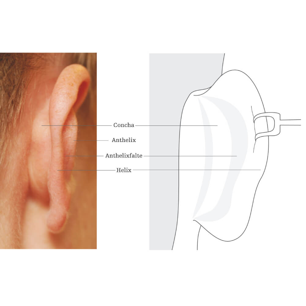
Anatomy for laypersons – for orientation

The concha is the cartilage visible from the front, right next to the ear canal (inner ear bowl); the helix is the soft outer rim of the ear that normally curves straight back, and the antihelix is the outer edge of the concha.

From an anatomical perspective, there are three possible reasons why ears may appear to protrude:

  • an overly wide concha (inner ear bowl)
  • a blunt antihelix angle
  • a combination of both causes

Below is a schematic representation of the three variations.
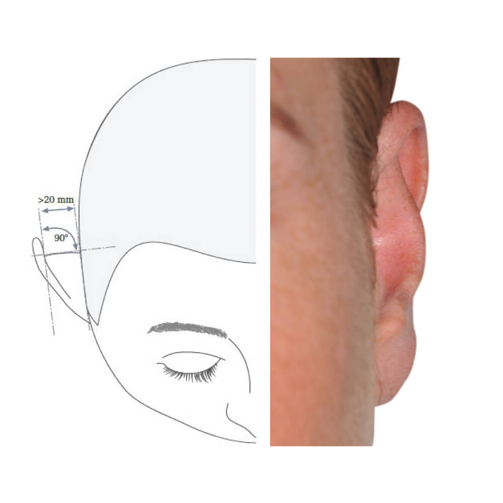
Normal, inconspicuous ear

Above is a depiction of a typical, inconspicuous ear: the helix runs parallel to the skull and forms a 90° angle with the antihelix. The distance between the skull and the helix is approximately 2 cm, corresponding to a normally wide concha.
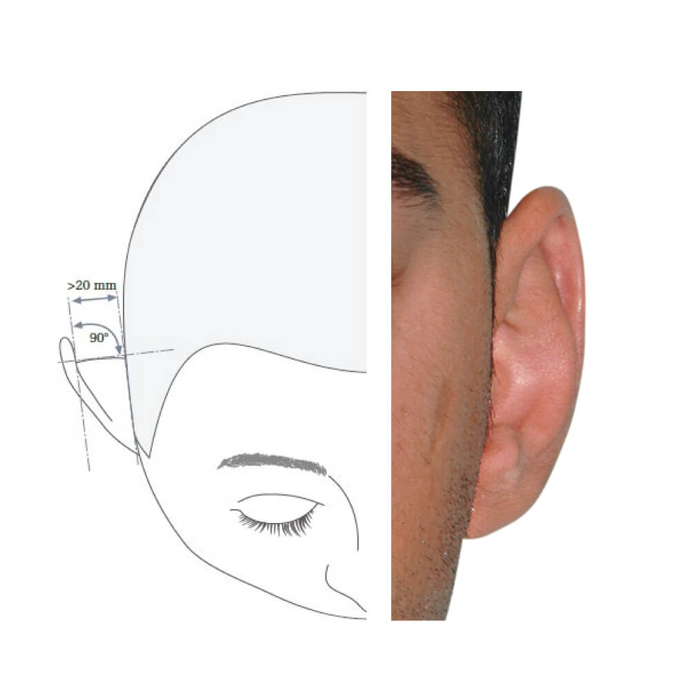
Below is an ear with a blunt antihelix angle but a normally wide concha

You can see the upper part of the ear protruding outward, while the inner ear bowl (concha) is of normal width.
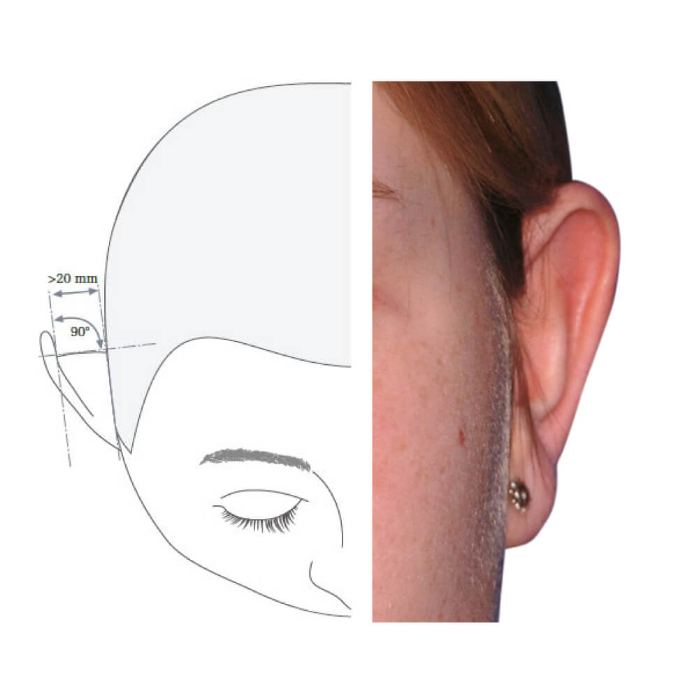
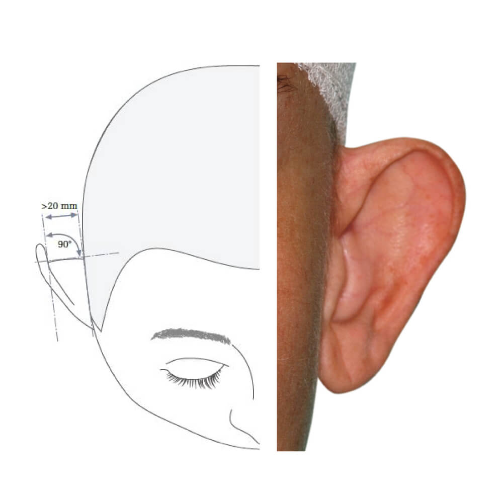
Below is an ear with a particularly wide concha but a normal antihelix angle

It is clearly visible how wide the inner ear bowl (concha) is, while the helix angles back normally (90°).
Below is an ear with a combination of both causes that can make an ear appear to protrude

Wide concha (inner ear bowl) and a blunt antihelix angle—here, the helix protrudes at a blunt angle and is visible from the front.

You can see that the inner ear bowl is wide, and the antihelix is barely visible due to the particularly blunt antihelix angle (arrow).

Interestingly, the most common form is a combination: an overly wide concha combined with a blunt antihelix angle.

For a beautiful result, the following details often need to be considered:

  • If the earlobes also protrude, they must be corrected through a separate, additional procedure (earlobe correction).
  • If cartilage segments along the antihelix fold are prominent, they should also be corrected (for example: protruding antitragus).

There are two surgical principles.

SUTURE METHOD

(I consider this a poor technique and therefore never use it)
The ear cartilage is bent into the desired position using shaping sutures (suture method); however, this method never takes into account whether the concha is too wide. It is simply pulled back to make it appear narrower— the same is done with the helix.

The suture method is fundamentally not a good approach because the cartilage is only “deformed” rather than “reshaped” (i.e., possibly narrowed). Additionally, the cartilage almost always remains under tension, which often causes it to return to its original position and the ear to protrude again.

SCALPEL METHOD

With a scalpel, any desired shape change can be made. The surgical result is permanent, and there is no risk of recurrence because the ear cartilage does not remain under tension.

A precise analysis and careful planning of the procedure are essential for the operation’s success.
Before and After Photos

All images on this website are of patients who were operated on by me. They have not been edited or altered in any way.

Ear Correction Overview

Below is a summary of the necessary steps and preparations associated with the ear correction procedure.

Initial Consultation

Initial Consultation

Have you already purchased the book “Ear Correction” for 40 euros at our clinic or online? Then the initial consultation on the topic of correcting protruding ears is included. A first consultation usually lasts around 30 minutes. Even if you have already had an aesthetic procedure performed at another clinic and are dissatisfied with the result, I am happy to advise you.
My personal tip for you: Always and as a general rule, seek a second opinion before undergoing any procedure (this is, of course, not included with the book purchase).

Surgery Preparation

Surgery Preparation

If the procedure is performed under local anesthesia

For hygiene regulations, a blood test (so-called “small blood panel”) is required, including HIV and Hepatitis A, B, and C. If you are socially insured in Austria and the procedure is medically indicated, there are no additional costs for the blood test.

If the procedure is performed under general anesthesia

Pre-operative preparation includes a blood test (so-called “large blood panel”), a chest X-ray from the age of 50, and surgical clearance by a general practitioner or internist.

In children, clearance is provided by a pediatrician and the chest X-ray may be omitted. If you are socially insured in Austria and the procedure is medically indicated, there are no additional costs in a public hospital. The same applies to surgical clearance by an internist or general practitioner who has a contract with the standard Austrian social insurance providers. For private doctors, the insurance covers about 50% of the costs.

However, my clinic does not have contracts with the social insurance institutions, which means the procedure is generally carried out privately and must therefore be paid for, unless the patient is under 16 years old and has private insurance.

Hospital Stay

Hospital Stay

Outpatient or day clinic (no overnight stay)

Anesthesia

Anesthesia

The procedure is usually performed under local anesthesia, so there are no additional costs, as no anesthesiologist is required for the surgery. If the procedure is performed under general anesthesia, an anesthesiologist must be present. The anesthesiologist’s fee varies depending on the duration of the surgery, ranging from €450 to €1200. On average, you should expect €600 to €700.

Costs

Costs

Depending on what needs to be corrected in addition to the protruding ears (earlobes, protruding cartilage sections, etc.), the surgical fee ranges from €2,800 to €4,500.

Rounds & Follow-up Appointments

Rounds & Follow-up Appointments

Are included in the surgical fee.

Is the correction of protruding ears technically difficult?

Ear correction is a fairly demanding procedure that should not be underestimated. It is important not to follow a standard approach but to also consider additional, often overlooked details such as a prominent antitragus or protruding earlobes when planning the surgery.

What surgical methods are available?

There are two main surgical techniques: either the ear cartilage is bent into the desired position using shaping sutures (suture method), or it is sculpted with a scalpel. The suture method is not a good approach, as the cartilage is only “deformed” rather than “reshaped.” Additionally, the cartilage almost always remains under tension, which often leads to it returning to its original position and the ear protruding again. In contrast, using a scalpel allows any desired shape change to be made, and the surgical result is permanent.

What is the suture method worth?

The suture method is neither modern nor effective and is therefore not recommended.

Where are the scars located after ear correction?

The incision is made behind the ear, so the scars are not visible.

From what age can children undergo surgery?

The procedure should ideally be performed under local anesthesia, which is usually possible from the age of 8. If correction is desired earlier, general anesthesia is required.

Does ear correction interfere with ear growth?

By the age of six, ears have reached 85% of their length and 95% of their width. Beyond that, surgically corrected ears continue to grow normally, so children can be safely operated on.

Is the procedure outpatient?

If performed under local anesthesia, the hospital can always be left on the same day. This is usually also possible with general anesthesia if the operation is done in the morning.

Does health insurance cover the cost of the procedure?

In Austria, health insurance covers the cost of ear correction up to the age of 16.

What complications can occur?

Medical complications such as bleeding, infections, or skin necrosis are extremely rare. The most common complications relate to the result: the ears protrude again after some time, the ears are asymmetrical, or the earlobes were not corrected, etc. Aesthetically unsatisfactory results can almost always be corrected.

What are the long-term results after ear correction?

If performed correctly and successfully, the long-term results are excellent. If the suture method was used, there is a relatively high chance that the ears will protrude again after some time.